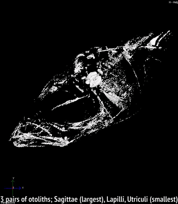
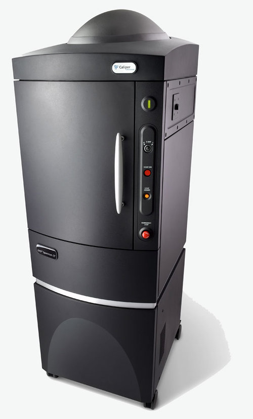
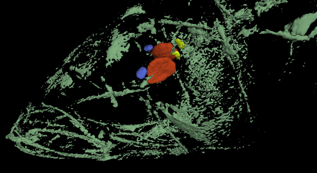
26 March 2018. Today we got our first glimpse of an incredible new way of imaging the inner calcified structures of a fish body, particularly the ear bones (otoliths), of which every teleost fish has six, three on each side inside the skull. Otoliths are long known to fish ecologists for their properties to record and store information about a fish’s age, growth and habitat. With an adult Atlantic silverside, Hannes visited John Shepherd, facilities scientist and member of the Goldhamer lab at UConn, Storrs (Biology Physics Building), who showed us the use of a new, state-of-the-art micro CT-scanner (IVIS). Turns out, the system effortlessly imaged all six otoliths inside of the fish’s head. Later in the year, we will use the technique to image silversides reared at contrasting CO2 conditions to see whether they differ in their otolith size, volume, and structure. Thank you, John, for this truly inspiring demonstration!