Web cookies (also called HTTP cookies, browser cookies, or simply cookies) are small pieces of data that websites store on your device (computer, phone, etc.) through your web browser. They are used to remember information about you and your interactions with the site.
Purpose of Cookies:
Session Management:
Keeping you logged in
Remembering items in a shopping cart
Saving language or theme preferences
Personalization:
Tailoring content or ads based on your previous activity
Tracking & Analytics:
Monitoring browsing behavior for analytics or marketing purposes
Types of Cookies:
Session Cookies:
Temporary; deleted when you close your browser
Used for things like keeping you logged in during a single session
Persistent Cookies:
Stored on your device until they expire or are manually deleted
Used for remembering login credentials, settings, etc.
First-Party Cookies:
Set by the website you're visiting directly
Third-Party Cookies:
Set by other domains (usually advertisers) embedded in the website
Commonly used for tracking across multiple sites
Authentication cookies are a special type of web cookie used to identify and verify a user after they log in to a website or web application.
What They Do:
Once you log in to a site, the server creates an authentication cookie and sends it to your browser. This cookie:
Proves to the website that you're logged in
Prevents you from having to log in again on every page you visit
Can persist across sessions if you select "Remember me"
What's Inside an Authentication Cookie?
Typically, it contains:
A unique session ID (not your actual password)
Optional metadata (e.g., expiration time, security flags)
Analytics cookies are cookies used to collect data about how visitors interact with a website. Their primary purpose is to help website owners understand and improve user experience by analyzing things like:
How users navigate the site
Which pages are most/least visited
How long users stay on each page
What device, browser, or location the user is from
What They Track:
Some examples of data analytics cookies may collect:
Page views and time spent on pages
Click paths (how users move from page to page)
Bounce rate (users who leave without interacting)
User demographics (location, language, device)
Referring websites (how users arrived at the site)
Here’s how you can disable cookies in common browsers:
1. Google Chrome
Open Chrome and click the three vertical dots in the top-right corner.
Go to Settings > Privacy and security > Cookies and other site data.
Choose your preferred option:
Block all cookies (not recommended, can break most websites).
Block third-party cookies (can block ads and tracking cookies).
2. Mozilla Firefox
Open Firefox and click the three horizontal lines in the top-right corner.
Go to Settings > Privacy & Security.
Under the Enhanced Tracking Protection section, choose Strict to block most cookies or Custom to manually choose which cookies to block.
3. Safari
Open Safari and click Safari in the top-left corner of the screen.
Go to Preferences > Privacy.
Check Block all cookies to stop all cookies, or select options to block third-party cookies.
4. Microsoft Edge
Open Edge and click the three horizontal dots in the top-right corner.
Go to Settings > Privacy, search, and services > Cookies and site permissions.
Select your cookie settings from there, including blocking all cookies or blocking third-party cookies.
5. On Mobile (iOS/Android)
For Safari on iOS: Go to Settings > Safari > Privacy & Security > Block All Cookies.
For Chrome on Android: Open the app, tap the three dots, go to Settings > Privacy and security > Cookies.
Be Aware:
Disabling cookies can make your online experience more difficult. Some websites may not load properly, or you may be logged out frequently. Also, certain features may not work as expected.
29 April 2020. Despite the lockdown and the virus, this is a joyful day for the Baumann lab – an unassumingly delivered note from the Provost – and both the end of a chapter and the beginning of a new era of Life and Science.
I’m grateful to so many people who aided this path along the way. Zosia, the boys, family, friends, and the many scientific mentors along the way.
This is a truly good day, not belittling the crisis & death all around us.
20 March 2020. We are happy to announce that the prestigious journal Fish & Fisheries just published a comprehensive review about the role of sand lance in the Northwest Atlantic Shelf ecosystem. The article, which came out of a workshop on this topic three years ago, reviews the the current state of knowledge about these enigmatic and important forage fish and urges continued efforts to better understand their role in the ecosystem and sensitivity to climate stressors.
This work represents the first comprehensive assessment of this important forage fish in the Northwest Atlantic, though similar efforts have been carried out in the Pacific Northwest and Europe. In the Atlantic, sand lance are observed to be a significant food source for the federally endangered Roseate tern, Atlantic sturgeon and cod, Harbor and Grey seals and Minke and Humpback whales. “This paper is a call to our peers and colleagues that there is a big gap in knowledge, and to bring more attention to these species as unmanaged forage fish,” says Staudinger.
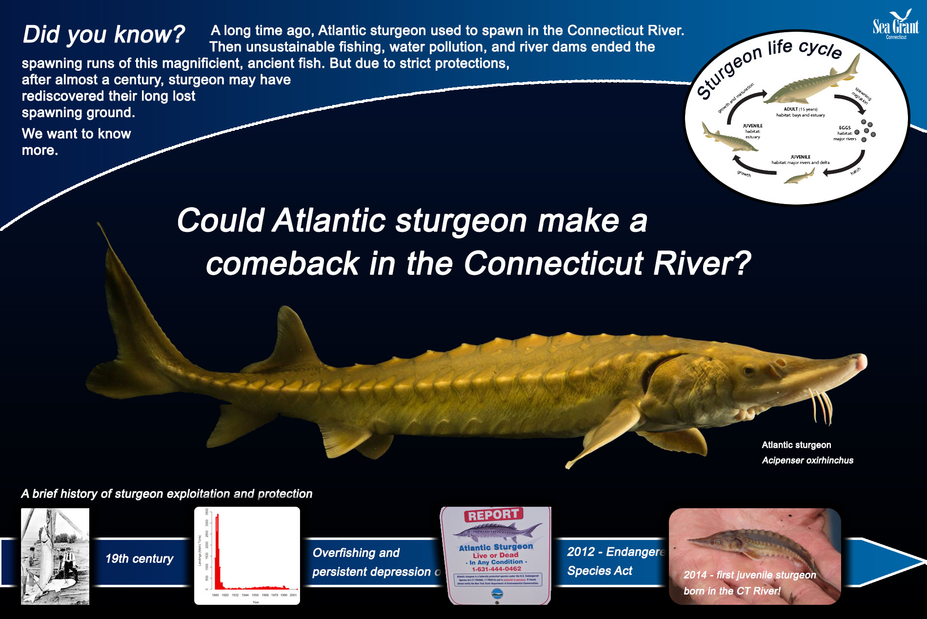
1 Feb 2020. We are elated to announce that Connecticut Sea Grant has decided to fund our latest research proposal to study Atlantic sturgeon in Long Island Sound and the Connecticut River! The project is funded for two years under the most recent Omnibus Funding call and will examine the growth and seasonal movement of these magnificent, ancient fish.
Kelli Mosca
The project will fund the Master thesis research of Kelli Mosca, the most recent addition to our lab! After receiving her Bachelors degree from the University of New Haven, Kelli became a dedicated seasonal worker at the Connecticut Department of Energy and Environmental Protection (CTDEEP), where she assisted particularly with the sturgeon monitoring program. This has made her the best possible graduate candidate to work this project. Welcome, Kelli!
Baumann, H., Savoy, T., Benway, J., and Pacileo, D. 2020. A re-emergent spawning population of Atlantic Sturgeon in the Connecticut River? Combined age analyses and telemetry data will provide new insights. Connecticut Sea Grant Program (NOAA) #R/LR-29, Feb 2020 - Feb 2022 ($150,000)
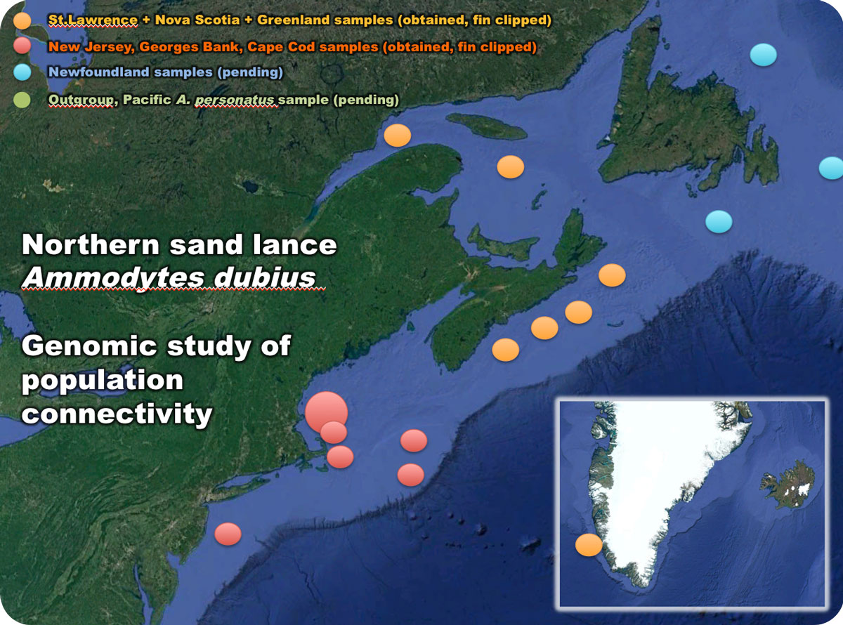
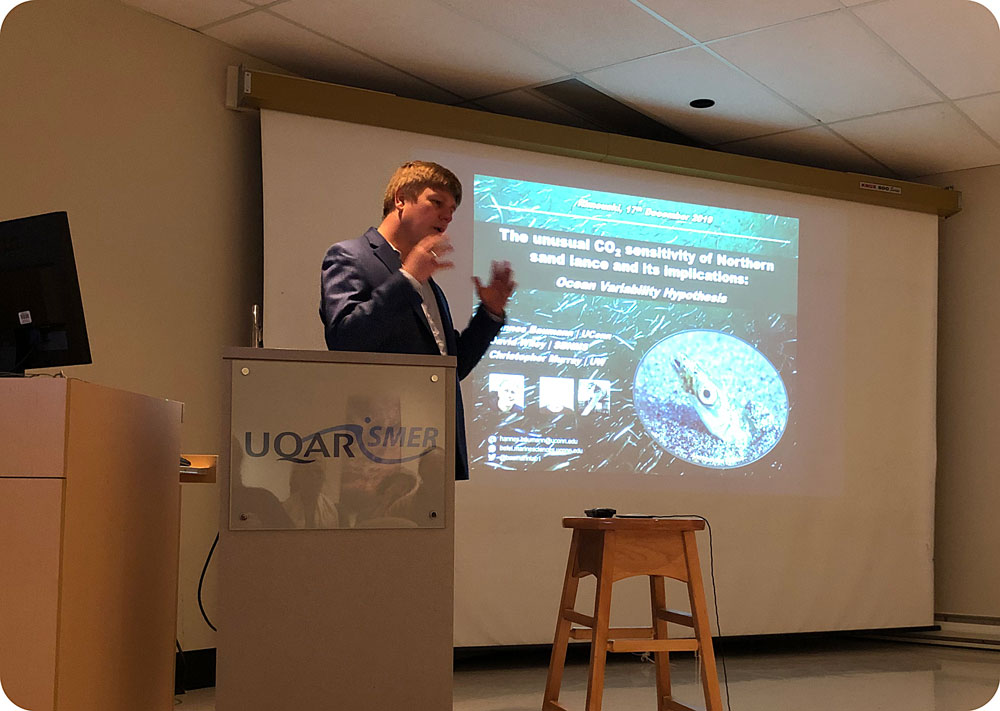
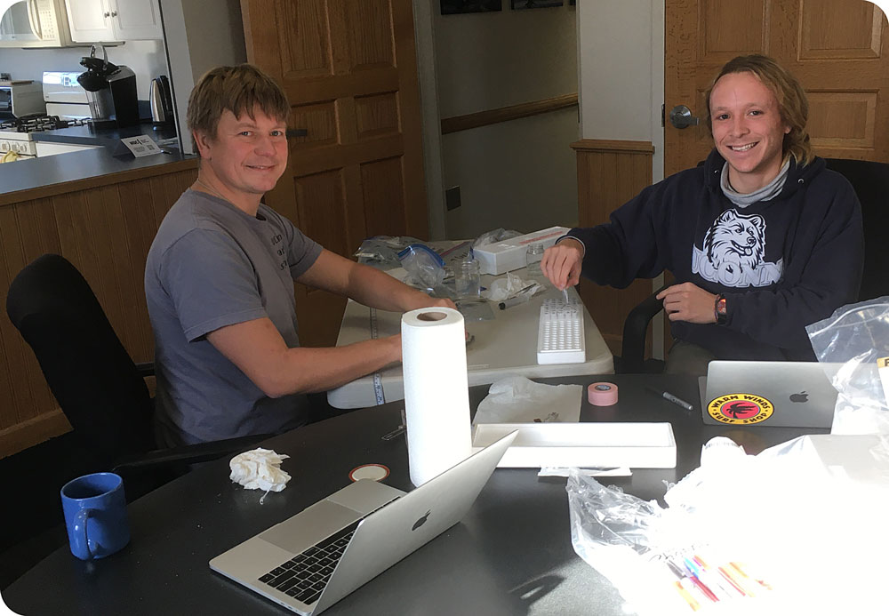
18 December 2019. Hannes and Lucas just returned from a spontaneous road trip to visit our good friends and collaborators at the University of Quebec in Rimouski (UQAR), Canada. We drove for over 10 hours (one-way) through snowstorms and icy voids to meet with Prof. Dominique Robert, who had collected sand lance samples from the Gulf of St. Lawrence and from Nova Scotia to be included in our new genomic study on the population connectivity of this species. Hannes gave a talk about our sand lance work and we saw a new institute in a new place, while frantically trying to stay warm amidst the brutal cold. Seeing the St. Lawrence in its icy, majestic beauty was a truly amazing experience.
Afterwards, we drove back through Maine and then repeated the fin-clipping of samples in Scituate at the Stellwagen Bank National Marine Sanctuary office, so we now have almost all samples in hand to start the DNA extraction and sequencing.
We are excited for the next steps!
Sand lance samples to be included in the genomic study
Rimouski on the south shore of the mighty and icy St. Lawrence River on 12/13/19
Lucas and Hannes listened to Corinne Burns talking about her PhD research at UQAR on 16 Dec 2019
Hannes gave a talk about sand lance research at UConn
The icy beauty of the St. Lawrence River
Sun glistening on the ice on the banks of the St. Lawrence River on 16 Dec 2019
Snow storm on the I91 in Vermont on 15 Dec 2019
Driving back through Maine on 17 Dec 2019 ... 6h of snowstorm
Sand lance sample thawing to be fin-clipped
Hannes and Lucas fin-clipping specimens in the Stellwagen Bank NMS office in Scituate on 18 Dec 2019
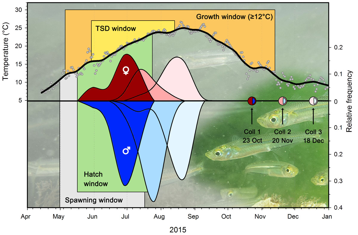
12 December 2019. We are happy to announce that Marine Ecology Progress Series just published our latest paper on Atlantic silversides, but this time not an experimental but a field study! During her time in our lab, Julie Pringle investigated the otolith microstructure of young-of-year silversides, finding intriguing patterns about differential growth in males and females that likely result in sex-selective survival during their growing season. Congratulations, Julie, well done!
This graph shows reconstructed hatch distributions of male and female Atlantic silversides sampled in fall 2015. Counting daily otolith increments, young-of-year fish caught in October could be reliably aged, whereas those from November and December where likely underaged because water temperatures had already decreased below their growth threshold. This graph compbines previous knowledge, environmental monitoring and results of otolith microstructure analysis.
From the abstract:
“We examined the utility of otolith microstructure analysis in young-of-year (YoY) Atlantic silversides Menidia menidia, an important annual forage fish species along the North American Atlantic coast. We first compared the known hatch window of a local population (Long Island Sound, USA) to otolith-derived hatch distributions, finding that YoY collected in October were reliably aged whereas survivors from November and December were progressively under- aged, likely due to the onset of winter ring formation. In all collections, males outnumbered fe- males, and both sexes had bimodal size distributions. However, while small and large females were almost evenly represented (~60 and ~40%, respectively), over 94% of all males belonged to the small size group. We then examined increment widths as proxies for somatic growth, which suggested that bimodal size distributions resulted from 2 distinct slow- and fast-growing YoY phe- notypes. Length back-calculations of October YoY confirmed this, because fast- and slow-growing phenotypes arose within common bi-weekly hatch intervals. We concluded that the partial sexual size dimorphism in this population resulted largely from sex-specific growth differences and not primarily from earlier female than male hatch dates, as predicted by the well-studied phenome- non of temperature-dependent sex determination (TSD) in this species. Furthermore, observed sex ratios were considerably less male-biased than reconstructed thermal histories and published laboratory TSD values predicted. Assuming that selective mortality is generally biased against slower growing individuals, this process would predominantly remove male silversides from the population and explain the more balanced sex ratios at the end of the growing season.”
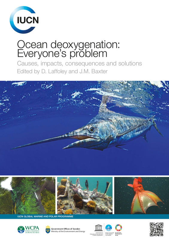
9 December 2019. During the COP25 summit in Madrid, the International Union for Conservation of Nature (IUCN) released its latest comprehensive report titled “Ocean deoxygenation: everyone’s problem” that compiles the current evidence for the ongoing, man-made decline in the oceans oxygen levels. The 588 page, 11 chapter wake-up call to these detrimental changes was produced by leading experts in the field. We are happy to announce that Hannes is one of the many authors of this document, co-authoring chapter 6 “Multiple stressors – forces that combine to worsen deoxygenation and its effects“.
From the executive summary:
“The equilibrium state of the ocean-atmosphere system has been perturbed these last few decades with the ocean becoming a source of oxygen for the atmosphere even though its oxygen inventory is only ~0.6% of that of the atmosphere. Different analyses conclude that the global ocean oxygen content has decreased by 1-2% since the middle of the 20th century. Global warming is expected to have contributed to this decrease, directly because the solubility of oxygen in warmer waters decreases, and indirectly through changes in the physical and biogeochemical dynamics.”
From the summary of chapter 6:
Human activities have altered not only the oxygen content of the coastal and open ocean, but also a variety of other physical, chemical and biological conditions that can have negative effects on physiological and ecological processes. As a result, marine systems are under intense and increasing pressure from multiple stressors.
The combined effects of ‘stressors’ can be greater than, less than, or different from the sum of each stressor alone, and there are large uncertainties surrounding their combined effects.
Warming, acidification, disease, and fisheries mortality are important common stressors that can have negative effects in combination with low oxygen.
Warming, deoxygenation, and acidification commonly co-occur because they share common causes. Increasing carbon dioxide (CO2) emissions simultaneously warm, deoxygenate, and acidify marine systems, and nutrient pollution increases the severity of deoxygenation and acidification.
A better understanding of the effects of multiple stressors on ocean ecosystems should improve the development of effective strategies to reduce the problem of deoxygenation and aid in identifying adaptive strategies to protect species and processes threatened by oxygen decline.
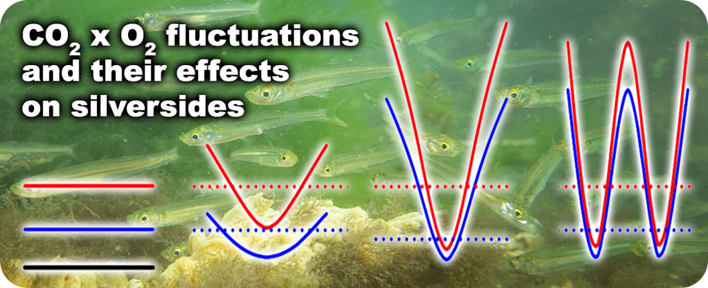
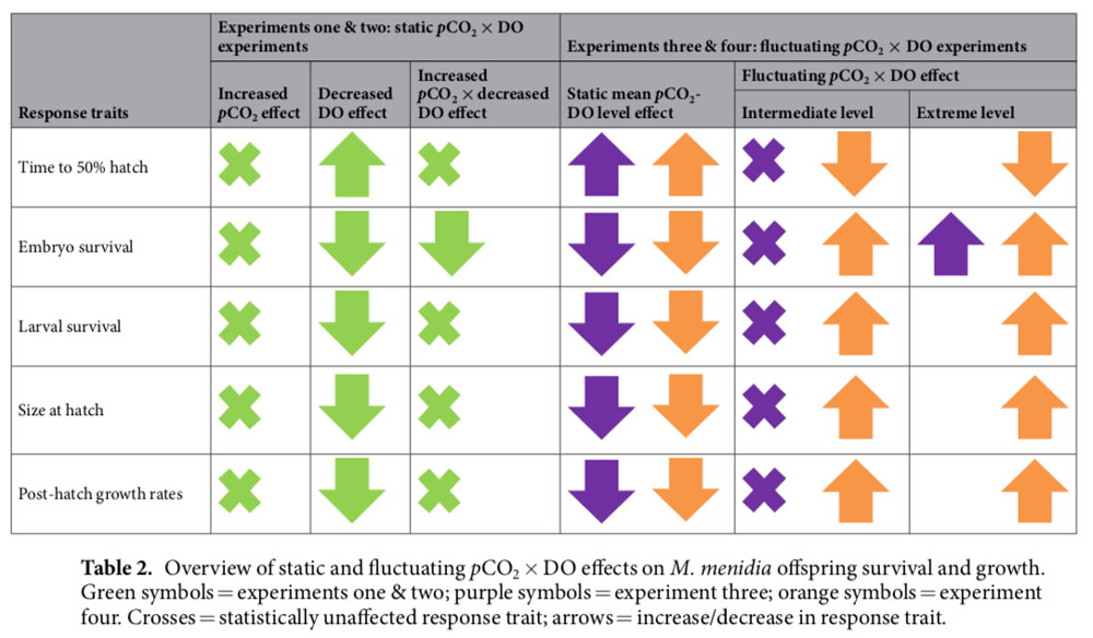
3 December 2019. We are happy and proud to share that Scientific Reports has published our latest research on the effects of fluctuating CO2 × O2 environments on the early life stages of Atlantic Silversides. The paper synthesizes findings of two years and four separate experiments – all conducted in our automated larval fish rearing system – to answer the question how current and future diel and tidal fluctuations in CO2 and O2 affect the survival and growth of silverside embryos and larvae.
The paper is a great demonstration of the vast capabilities of our system to simulate non-static conditions, which is a frontier in climate change research. Congrats to Emma Cross for pulling all the complex data together!
“Static low DO conditions severely decreased embryo survival, larval survival, time to 50% hatch, size at hatch and post-larval growth rates. Static elevated pco2 did not affect most response traits, however, a synergistic negative effect did occur on embryo survival under hypoxic conditions (3.0 mg L−1). Cycling CO2 × DO, however, reduced these negative effects of static conditions on all response traits with the magnitude of fluctuations influencing the extent of this reduction. This indicates that fluctuations in pco2 and DO may benefit coastal organisms by providing periodic physiological refuge from stressful conditions, which could promote species adaptability to climate change.”
The source data for this publication are openly available (and citable) from the BCO-DMO database. Head to Products -> Research Data to access them!
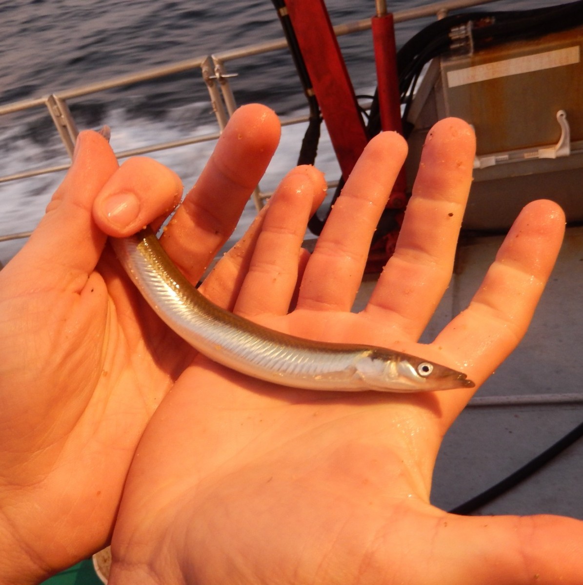
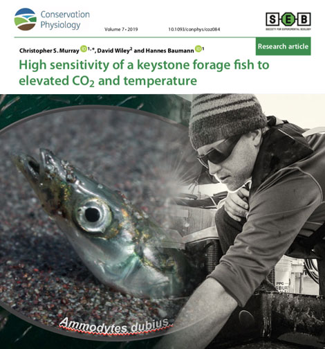
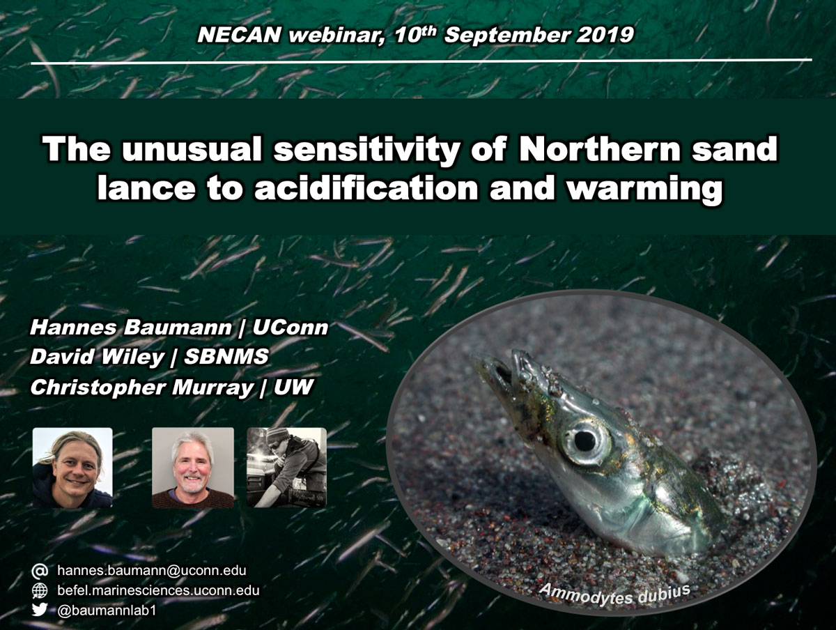
21 November 2019. We are excited to announce the Chris Murray‘s paper on the unusual, high sensitivity of early life Northern sand lance to acidification and warming has just been published in the journal of Conservation Physiology! This is the first publication of our extensive work on this enigmatic species.
Sand lance species play a key ecological role in most temperate to polar shelf ecosystems of the northern hemisphere, but they have remained unstudied with respect to their sensitivity to predicted future CO2 levels in the ocean. For the past three years (2016 – 2018), we have sampled and spawned with northern sand lance (Ammodytes dubius) from Stellwagen Bank National Marine Sanctuary and subsequently reared their embryos under factorial CO2 x temperature conditions to hatch and early larval stages. Our results were striking, in all years, high CO2 conditions severely reduced embryo survival up to 20-fold over controls, with strong synergistic reductions under combined high CO2 and temperature conditions. High CO2 also delayed hatching, reduced remaining endogenous energy reserves at hatch, and in combination with higher temperatures, reduced embryonic growth.
Indeed, given the observed effect sizes, northern sand lance might be the most CO2 sensitive fish species tested to date.
10 September 2019. Hannes started of the new 2019 NECAN Sea Grant Webinar Series with a presentation of our past years of research on the sensitivity of Northern sand lance (Ammodytes dubius) to ocean acidification and warming. The purpose of this webinar series is to highlight four projects funded through NOAA Sea Grant following the release of the NECAN paper published in Oceanography Magazine in 2015, “Ocean and Coastal Acidification off New England and Nova Scotia.”
Thanks to the more than 50 people who attended the webinar. If you have missed it, it’s accessible for free online. See below.
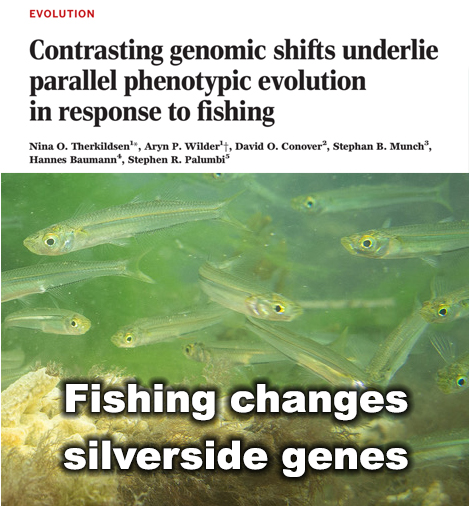
1 August 2019. We are overjoyed that our paper on genetic changes in experimental silverside populations subjected to strong size-selective fishing has just been published by Science!
Over recent decades, many commercially harvested fish have grown slower and matured earlier, which can translate into lower yields. Scientists have long suspected that rapid evolutionary change in fish caused by intense harvest pressure is the culprit.
Now, for the first time, researchers have unraveled genome-wide changes that prompted by fisheries – changes that previously had been invisible, according to a study published in Science by a team of researchers including Hannes Baumann, UConn assistant professor of Marine Sciences, who collaborated with researchers at Cornell University, the University of Oregon, the National Marine Fisheries Service, and Stanford University.
In unprecedented detail, the study shows sweeping genetic changes and how quickly those changes occur in fish populations extensively harvested by humans, says Baumann.
“Most people think of evolution as a very slow process that unfolds over millennial time scales, but evolution can, in fact, happen very quickly,” said lead author Nina Overgaard Therkildsen, Cornell assistant professor of conservation genomics in the Department of Natural Resources.
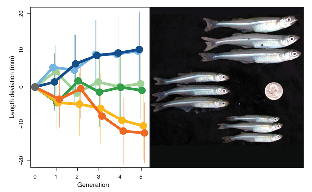
Observed shifts in adult size. Trends across generations in mean length at harvest (standardized as the difference from the mean of the control populations in each generation) ± the standard deviations in up-selected (blue shades), down-selected (yellow and orange shades), and control populations (green shades).
The all-pervasive human meddling in our planet’s affairs undeniably reached the genetic make-up of its organisms.
— Hannes Baumann, UConn.
In heavily exploited fish stocks, fishing almost always targets the largest individuals. “Slower-growing fish will be smaller and escape the nets better, thereby having a higher chance of passing their genes on to the next generations. This way, fishing can cause rapid evolutionary change in growth rates and other traits,” said Therkildsen. “We see many indications of this effect in wild fish stocks, but no one has known what the underlying genetic changes were.”
Therkildsen and her colleagues took advantage of an influential experiment published back in 2002. Six populations of Atlantic silversides, a fish that grows no bigger than 6 inches in length, had been subjected to intense harvesting in the lab. In two populations, the largest individuals were removed; in another two populations, the smallest individuals were removed; and in the final two populations, the fishing was random with respect to size.
After only four generations, these different harvest regimes had led to evolution of an almost two-fold difference in adult size between the groups. Therkildsen and her team sequenced the full genome of almost 900 of these fish to examine the DNA-level changes responsible for these striking shifts.
The team identified hundreds of different genes across the genome that changed consistently between populations selected for fast and slow growth. They also observed large linked-blocks of genes that changed in concert, dramatically shifting the frequencies of hundreds of genes all at the same time.
Surprisingly, these large shifts only happened in some of the populations, according to the new paper. This means that there were multiple genomic solutions for the fish in this experiment to get either larger or smaller.
“Some of these changes are easier to reverse than others, so to predict the impacts of fisheries-induced evolution, it is not enough to track growth rates alone, we need to monitor changes at the genomic level,” said Therkildsen.
When the experiment was originally conducted nearly two decades ago by co-authors David Conover, professor of biology at the University of Oregon, and Stephan Munch of the National Marine Fisheries Service, the tools to study the genomic basis of the rapid fisheries-induced evolution they observed were not available. Fortunately, Conover and Munch had the foresight to store the samples in a freezer, making it possible to now return – armed with modern DNA sequencing tools – and reveal the underlying genomic shifts.
Research like this can assess human impacts, and improve humanity’s understanding of “the speed, consequences and reversibility of complex adaptations as we continue to sculpt the evolutionary trajectories of the species around us,” Therkildsen said.
“What’s most fascinating about this is that life can find different genetic ways to achieve the same result. In this study, two experimental populations evolved smaller body size in response to the selective removal of the largest fish, which is what most trawl fisheries do. However, only by looking at the genetic level we demonstrated that these two experimental populations evolved via two completely different genetic paths,” says Baumann.
The good news for the Atlantic silversides is that the fisheries selection was able to tap into the large reservoir of genetic variation that exists across the natural range of this species from Florida into Canada, said Therkildsen: “That genetic bank fueled rapid adaptation in the face of strong fishing pressure. Similar responses may occur in response to climate-induced shifts in other species with large genetic variability.”
“Scientists have coined the term Anthropocene in recognition of the all-pervasive human alteration of the earth’s climate, oceans, and land. No matter how ‘pristine’ a piece of nature may look to us at first glance, examine it thoroughly enough and you will find a trace of human in it. Take a cup of water from the middle of Pacific Ocean and a handful of sand from a ‘pristine’ beach – and you will find little plastic particles under the microscope,” says Baumann. “The parallel to this study is that the all-pervasive human meddling in our planet’s affairs now undeniably reached the genetic make-up of its organisms. Today’s fishes may superficially look the same as always, but their genes are not. They bear witness to human alteration.”
In addition to Baumann, Therkildsen, Conover, and Munch, co-authors included former Cornell postdoctoral researcher Aryn P. Wilder, now a researcher at San Diego Zoo Institute for Conservation Research; and Stephen R. Palumbi, Stanford University.
This work was funded by the National Science Foundation.