Web cookies (also called HTTP cookies, browser cookies, or simply cookies) are small pieces of data that websites store on your device (computer, phone, etc.) through your web browser. They are used to remember information about you and your interactions with the site.
Purpose of Cookies:
Session Management:
Keeping you logged in
Remembering items in a shopping cart
Saving language or theme preferences
Personalization:
Tailoring content or ads based on your previous activity
Tracking & Analytics:
Monitoring browsing behavior for analytics or marketing purposes
Types of Cookies:
Session Cookies:
Temporary; deleted when you close your browser
Used for things like keeping you logged in during a single session
Persistent Cookies:
Stored on your device until they expire or are manually deleted
Used for remembering login credentials, settings, etc.
First-Party Cookies:
Set by the website you're visiting directly
Third-Party Cookies:
Set by other domains (usually advertisers) embedded in the website
Commonly used for tracking across multiple sites
Authentication cookies are a special type of web cookie used to identify and verify a user after they log in to a website or web application.
What They Do:
Once you log in to a site, the server creates an authentication cookie and sends it to your browser. This cookie:
Proves to the website that you're logged in
Prevents you from having to log in again on every page you visit
Can persist across sessions if you select "Remember me"
What's Inside an Authentication Cookie?
Typically, it contains:
A unique session ID (not your actual password)
Optional metadata (e.g., expiration time, security flags)
Analytics cookies are cookies used to collect data about how visitors interact with a website. Their primary purpose is to help website owners understand and improve user experience by analyzing things like:
How users navigate the site
Which pages are most/least visited
How long users stay on each page
What device, browser, or location the user is from
What They Track:
Some examples of data analytics cookies may collect:
Page views and time spent on pages
Click paths (how users move from page to page)
Bounce rate (users who leave without interacting)
User demographics (location, language, device)
Referring websites (how users arrived at the site)
Here’s how you can disable cookies in common browsers:
1. Google Chrome
Open Chrome and click the three vertical dots in the top-right corner.
Go to Settings > Privacy and security > Cookies and other site data.
Choose your preferred option:
Block all cookies (not recommended, can break most websites).
Block third-party cookies (can block ads and tracking cookies).
2. Mozilla Firefox
Open Firefox and click the three horizontal lines in the top-right corner.
Go to Settings > Privacy & Security.
Under the Enhanced Tracking Protection section, choose Strict to block most cookies or Custom to manually choose which cookies to block.
3. Safari
Open Safari and click Safari in the top-left corner of the screen.
Go to Preferences > Privacy.
Check Block all cookies to stop all cookies, or select options to block third-party cookies.
4. Microsoft Edge
Open Edge and click the three horizontal dots in the top-right corner.
Go to Settings > Privacy, search, and services > Cookies and site permissions.
Select your cookie settings from there, including blocking all cookies or blocking third-party cookies.
5. On Mobile (iOS/Android)
For Safari on iOS: Go to Settings > Safari > Privacy & Security > Block All Cookies.
For Chrome on Android: Open the app, tap the three dots, go to Settings > Privacy and security > Cookies.
Be Aware:
Disabling cookies can make your online experience more difficult. Some websites may not load properly, or you may be logged out frequently. Also, certain features may not work as expected.
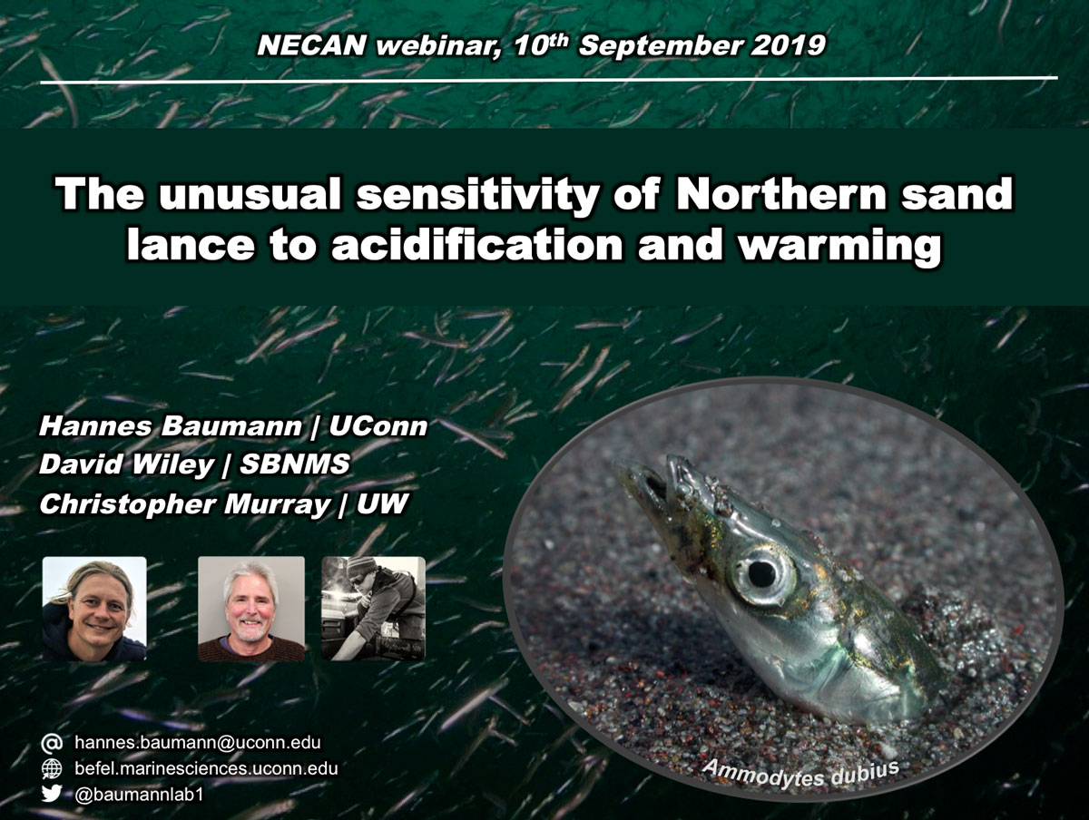
10 September 2019. Hannes started of the new 2019 NECAN Sea Grant Webinar Series with a presentation of our past years of research on the sensitivity of Northern sand lance (Ammodytes dubius) to ocean acidification and warming. The purpose of this webinar series is to highlight four projects funded through NOAA Sea Grant following the release of the NECAN paper published in Oceanography Magazine in 2015, “Ocean and Coastal Acidification off New England and Nova Scotia.”
Thanks to the more than 50 people who attended the webinar. If you have missed it, it’s accessible for free online. See below.
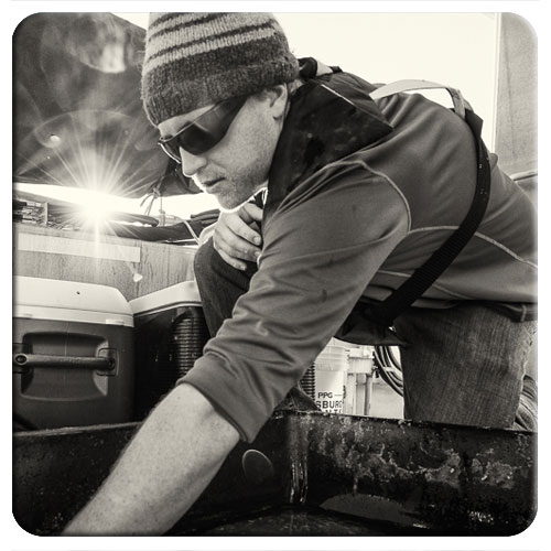
15 August 2019. The Baumann lab is happy to announce that Chris Murray has started his new chapter of life and science at the west coast with the University of Washington. Congrats Chris, we know you will do great!
Chris started his PhD at UConn/Avery Point in September 2014, after finishing his MS in May 2014 at Stony Brook University, NY. While building on his experience in ocean acidification research, for his PhD he studyied multi-stressor effects of OA and hypoxia on coastal marine fishes. He had an outstanding part in designing and building our factorial larval rearing system ("Larval city") in UConn Rankin Seawater lab. The system allows up to nine independent, static or fluctuating CO2 x O2 environments simultaneously. It has been in full use during spring and summer months of the past four years.
After a phenomenally dedicated four years, Chris defended his PhD in December 2018 and recently graduated with this PhD from UConn.
His thesis titled An experimental evaluation of the sensitivity of coastal marine fishers acidification, hypoxia, and warming
Murray, C.S.*, Wiley, D., and Baumann, H. High sensitivity of a keystone forage fish to elevated CO2 and temperature. Conservation Physiology (resubmitted)
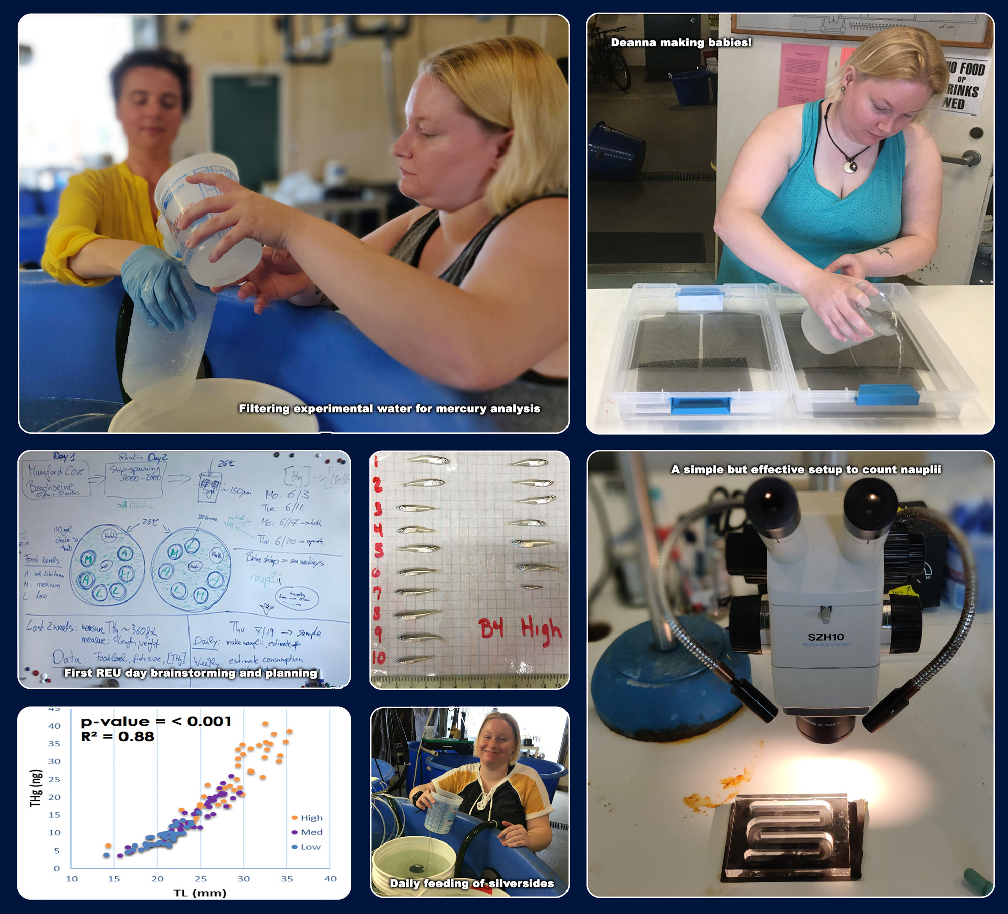
10 August 2019.Deanna Elliott from Arizona State University has just successfully completed her summer research project as our third NSF-REU student. For her REU-project she reared Atlantic silverside larvae under different feeding regimes to create fish of different body sizes and then analyzed them for trace levels of mercury in their tissue. She tested the hypothesis that mercury concentrations in fish can be used as a proxy for ingestion rates, which are important to trophic ecosystem models to perform better.
Here’s what Deanna had to say about her REU research experience:
This summer, I spent 10 weeks in the Baumann Evolutionary Fish Ecology lab and had a blast! The entire lab was incredibly welcoming, and made me feel at home immediately. We jumped right into my project and I had so many new experiences, it was almost overwhelming. We went seining for silversides in Mumford Cove, fertilized fish eggs… I became a Fish Mommy for the first time, rearing approximately 500 juvenile silversides for five weeks—I had never even had a fish tank before! I also got valuable experience in the chemistry lab, analyzing the mercury content of my Fish Babies. I felt very welcomed and received a lot of encouragement on my project and the presentation I had to give at the end of the program. Hannes and Zosia especially made me feel appreciated and supported, and that made all the difference in my experience with UCONN’s marine biology REU.
Check out some of the impressions from Deanna’s time at UConn. Great job, Deanna!
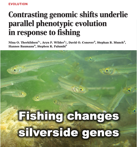
1 August 2019. We are overjoyed that our paper on genetic changes in experimental silverside populations subjected to strong size-selective fishing has just been published by Science!
Over recent decades, many commercially harvested fish have grown slower and matured earlier, which can translate into lower yields. Scientists have long suspected that rapid evolutionary change in fish caused by intense harvest pressure is the culprit.
Now, for the first time, researchers have unraveled genome-wide changes that prompted by fisheries – changes that previously had been invisible, according to a study published in Science by a team of researchers including Hannes Baumann, UConn assistant professor of Marine Sciences, who collaborated with researchers at Cornell University, the University of Oregon, the National Marine Fisheries Service, and Stanford University.
In unprecedented detail, the study shows sweeping genetic changes and how quickly those changes occur in fish populations extensively harvested by humans, says Baumann.
“Most people think of evolution as a very slow process that unfolds over millennial time scales, but evolution can, in fact, happen very quickly,” said lead author Nina Overgaard Therkildsen, Cornell assistant professor of conservation genomics in the Department of Natural Resources.
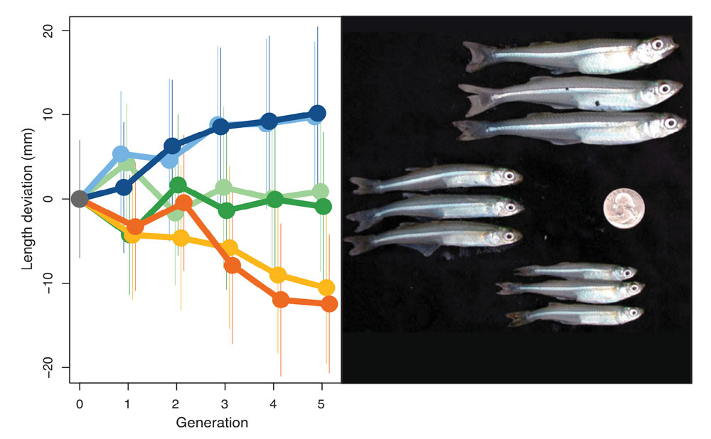
Observed shifts in adult size. Trends across generations in mean length at harvest (standardized as the difference from the mean of the control populations in each generation) ± the standard deviations in up-selected (blue shades), down-selected (yellow and orange shades), and control populations (green shades).
The all-pervasive human meddling in our planet’s affairs undeniably reached the genetic make-up of its organisms.
— Hannes Baumann, UConn.
In heavily exploited fish stocks, fishing almost always targets the largest individuals. “Slower-growing fish will be smaller and escape the nets better, thereby having a higher chance of passing their genes on to the next generations. This way, fishing can cause rapid evolutionary change in growth rates and other traits,” said Therkildsen. “We see many indications of this effect in wild fish stocks, but no one has known what the underlying genetic changes were.”
Therkildsen and her colleagues took advantage of an influential experiment published back in 2002. Six populations of Atlantic silversides, a fish that grows no bigger than 6 inches in length, had been subjected to intense harvesting in the lab. In two populations, the largest individuals were removed; in another two populations, the smallest individuals were removed; and in the final two populations, the fishing was random with respect to size.
After only four generations, these different harvest regimes had led to evolution of an almost two-fold difference in adult size between the groups. Therkildsen and her team sequenced the full genome of almost 900 of these fish to examine the DNA-level changes responsible for these striking shifts.
The team identified hundreds of different genes across the genome that changed consistently between populations selected for fast and slow growth. They also observed large linked-blocks of genes that changed in concert, dramatically shifting the frequencies of hundreds of genes all at the same time.
Surprisingly, these large shifts only happened in some of the populations, according to the new paper. This means that there were multiple genomic solutions for the fish in this experiment to get either larger or smaller.
“Some of these changes are easier to reverse than others, so to predict the impacts of fisheries-induced evolution, it is not enough to track growth rates alone, we need to monitor changes at the genomic level,” said Therkildsen.
When the experiment was originally conducted nearly two decades ago by co-authors David Conover, professor of biology at the University of Oregon, and Stephan Munch of the National Marine Fisheries Service, the tools to study the genomic basis of the rapid fisheries-induced evolution they observed were not available. Fortunately, Conover and Munch had the foresight to store the samples in a freezer, making it possible to now return – armed with modern DNA sequencing tools – and reveal the underlying genomic shifts.
Research like this can assess human impacts, and improve humanity’s understanding of “the speed, consequences and reversibility of complex adaptations as we continue to sculpt the evolutionary trajectories of the species around us,” Therkildsen said.
“What’s most fascinating about this is that life can find different genetic ways to achieve the same result. In this study, two experimental populations evolved smaller body size in response to the selective removal of the largest fish, which is what most trawl fisheries do. However, only by looking at the genetic level we demonstrated that these two experimental populations evolved via two completely different genetic paths,” says Baumann.
The good news for the Atlantic silversides is that the fisheries selection was able to tap into the large reservoir of genetic variation that exists across the natural range of this species from Florida into Canada, said Therkildsen: “That genetic bank fueled rapid adaptation in the face of strong fishing pressure. Similar responses may occur in response to climate-induced shifts in other species with large genetic variability.”
“Scientists have coined the term Anthropocene in recognition of the all-pervasive human alteration of the earth’s climate, oceans, and land. No matter how ‘pristine’ a piece of nature may look to us at first glance, examine it thoroughly enough and you will find a trace of human in it. Take a cup of water from the middle of Pacific Ocean and a handful of sand from a ‘pristine’ beach – and you will find little plastic particles under the microscope,” says Baumann. “The parallel to this study is that the all-pervasive human meddling in our planet’s affairs now undeniably reached the genetic make-up of its organisms. Today’s fishes may superficially look the same as always, but their genes are not. They bear witness to human alteration.”
In addition to Baumann, Therkildsen, Conover, and Munch, co-authors included former Cornell postdoctoral researcher Aryn P. Wilder, now a researcher at San Diego Zoo Institute for Conservation Research; and Stephen R. Palumbi, Stanford University.
This work was funded by the National Science Foundation.
Deanna Elliott is a junior at Arizona State University who has joined the Baumann lab in summer 2019 as our third NSF-REU student. Deanna has experimented with locusts before, but now strives to become an expert fish rearer. Her project will rear Atlantic silverside larvae under different feeding regimes to create fish of different body sizes and then analyze the these fish for trace levels of mercury in their tissue. She will test the hypothesis that mercury concentrations in fish can be used as a proxy for ingestion rates, which are important to improve trophic ecosystem models. Welcome, Deanna!
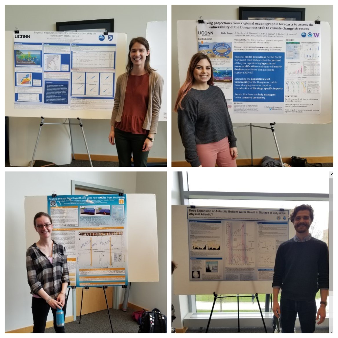
UConn DMS students present their research (u.l. Kelly McGarry, u.r. Halle Berger, l.l. Sarah McCart, l.r. Alec Shub
By Callie Concannon. On April 30th, four graduate students from the Marine Science Department traveled to UConn, Storrs to present their research at UConn’s 2nd Climate Research Symposium cohosted by the Geology and Marine Science departments. The students were Kelly McGarry (Ph.D student; top left), Halle Berger (Master’s student; top right), Sarah McCart (Master’s student; bottom left) and Alec Shub (Master’s student; bottom right). Everyone’s presentations were well received, and Sarah McCart even won the graduate student poster competition!
The event featured two keynote speakers; Professor Margaret Rubega of UConn, and Professor Tim Cronin of MIT. Professor Rubega talked about science communication and how the scientific community could better communicate their climate change research to non-scientists without using overbearing jargon and too many words. Professor Cronin gave a speech on his past research on the suppression of Arctic air formation with climate warming.
McCart, S., Lund, D., Seeley, E., Asimov, P., Lewis, M., and Mudahy, A.L. Testing the sea level hypothesis with new results from the Pacific.
McGarry, K., Siedlecki, S., Alin, S., and Salisbury, J. Empirical models for estimating the carbonate system along the northeastern coast of the U.S.
Berger, H., Siedlecki, S., Matassa, C., Alin, S., Kaplan, I., Pilcher, D., and Newton, J. Using projections from regional oceanographic forecasts to assess the vulnerability of the Dungeness crab to climate change stressors.
Shub, A., Lund, D., and Mudahy, A.L., Does expansion of Antarctic bottom water result in storage of CO2 in the abyssal Atlantic?
3 May 2019. It is Emma’s 30th birthday today, so naturally she celebrates it by starting a new, large experiment with Atlantic silversides, thus sharing her special day with more than 5,000 little embryos that are now developing in our system.
Like in our previous experiments, we are mimicking current and future coastal environments that fluctuate daily in CO2 and oxygen levels – thanks to our computer-controlled system that manipulates these levels in up to nine tanks simultaneously.
But this time, our additional goal is to keep track of sib-ship. We produced full sibs (same mother, same father), half-sibs (same mother or father, different father or mother) and unrelated individuals, and by keeping them separate we will later be able to calculate additive genetic variances in the various traits under different conditions (i.e., heritability) and examine trait correlations.
As usual, this could not be done by one person, so the entire lab helped preparing, seining, and fertilizing embryos on this frantic day. Great job all!
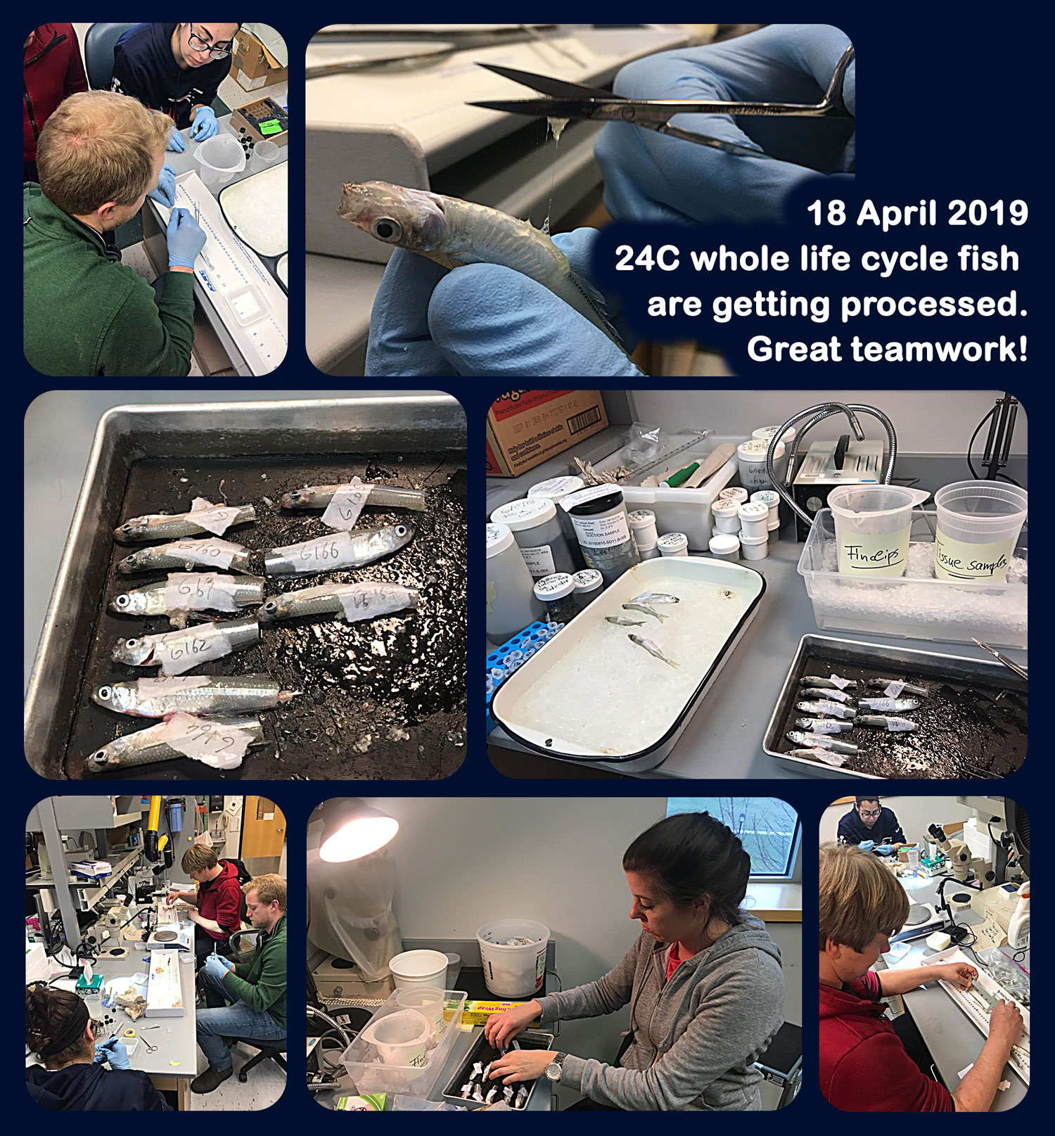
18 April 2019. This Thursday was a long day in the Baumann lab, because we sampled and processed over 200 adult silversides from a unique experiment. These fish were fertilized in the lab and reared from eggs to adulthood under different temperatures and contrasting CO2 conditions. We are interested to see, if future ocean conditions have measurable effects on this species fecundity, growth, and oocyte characteristics. We also took tissue and genetic samples, with an effective line-up of hands, i.e., Hannes, Emma, Chris, Callie and Lucas.
Good teamwork all!
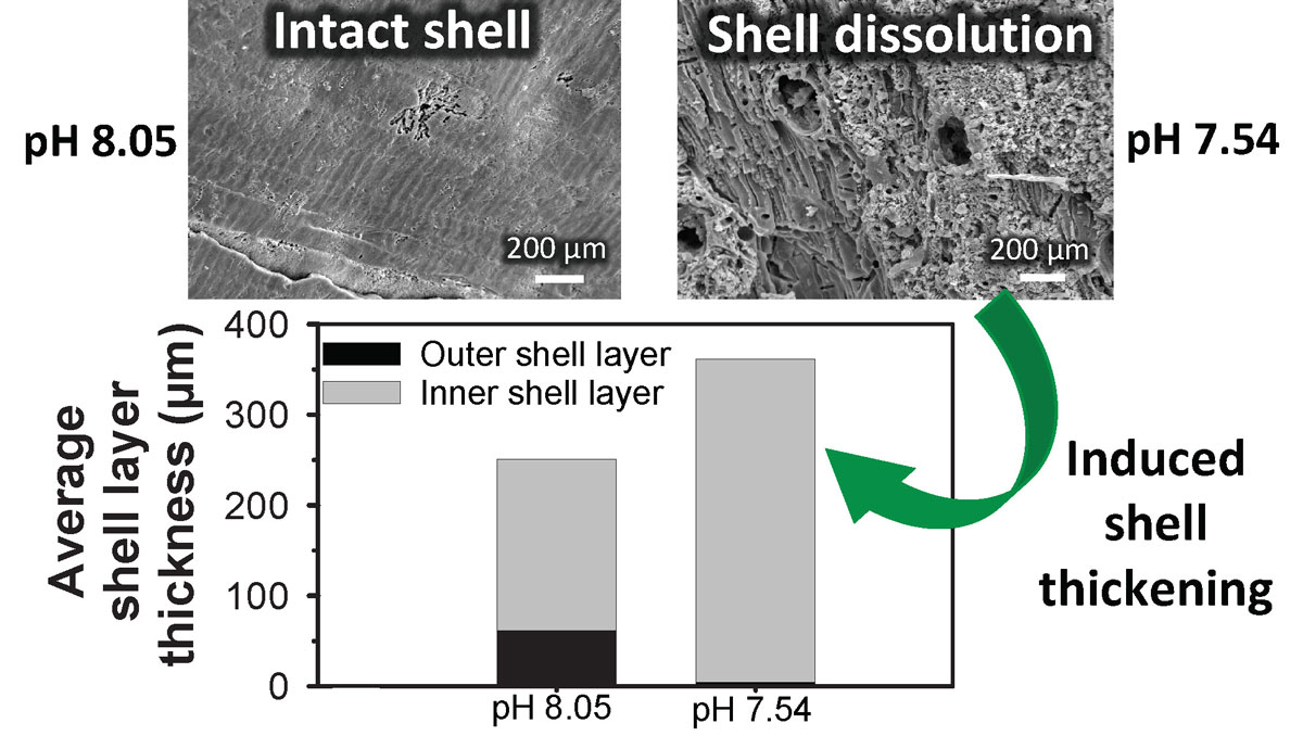
15 April 2019. Today, Emma is happy to report that Environmental Science & Technology have just published the latest paper from her PhD about brachiopod resilience to future ocean acidification. This project involved long-term culturing of a polar and a temperate brachiopod under future ocean acidification and warming conditions. Substantial shell dissolution posed a threat to both species under ocean acidification, with more extensive dissolution occurring in the polar species.
Unexpectedly, we discovered that brachiopods thicken their shell from the inner shell surface when extensive dissolution occurs at the outer shell surface under ocean acidification. This is an important finding to further our understanding of how predicted vulnerable marine calcifiers might cope under future environmental change.
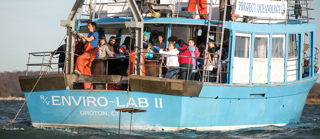
Project Oceanology students onboard the “Enviro-Lab II” retrieve a trawl in the Thames River Mouth (Photo: Anna Sawin)
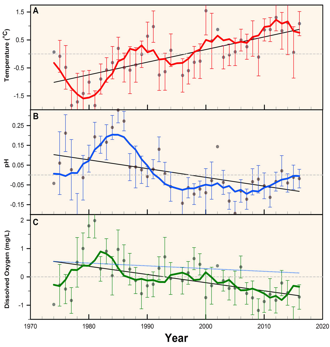
21 March 2019. We are happy to announce that Marine Environmental Research just published our most recent paper about long-term ecological change in eastern Long Island Sound based on data collected by Project Oceanology!For his Master’s thesis, Jacob Snyder painstakingly retrieved and digitized more than 40 years of environmental observations from Project Oceanology. This non-profit ocean literacy organization has educated middle and high school students on boat trips to nearby estuarine sites for decades. For the first time, his work allowed a quantitative evaluation of these data and glimpses into the abiotic and biotic changes in nearshore waters of Eastern Long Island Sound.
Highlights
Citizen-science observations revealed rapid warming, acidification, and dissolved oxygen loss over the past 40 years in eastern Long Island Sound
Otter trawl catches showed significant decreases in overall species diversity and richness
Cold-water adapted species (American lobster, winter flounder) decreased, but warm-water adapted species (spider crabs) increased since 1997